Ocular Revision
by Paul Vanouse, 2010
Vanouse
projects
VIDEO DOCUMENATION


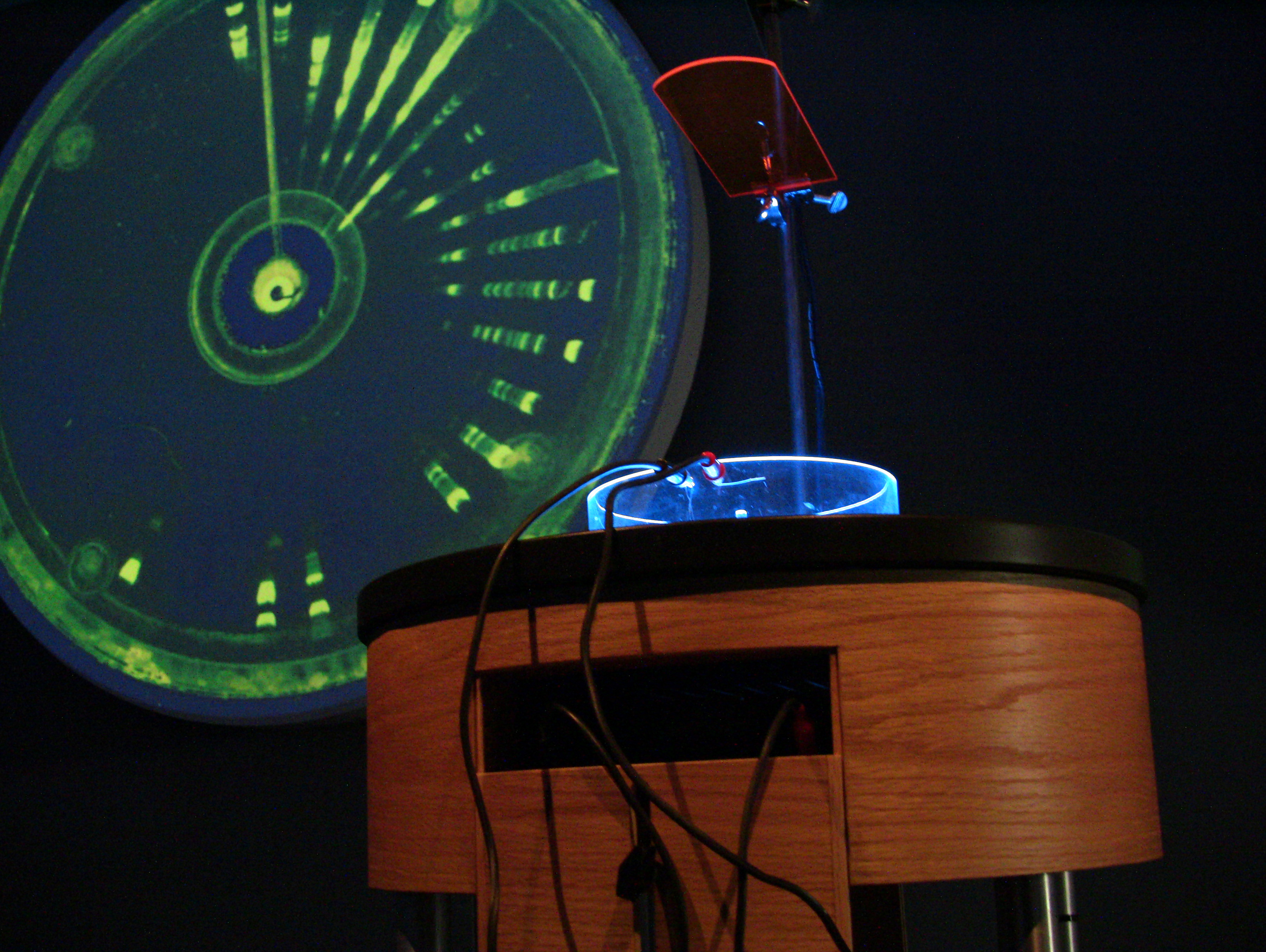
Ocular Revision, documentation of first imaging experiment.
Biology, which defines the cell as the basic unit of life, is a discipline broadly impacted by an ocular frenzy of the Modern period.  Prior to the microscope, the life sciences were limited to anatomical investigations.  A timeline of optical devices of this period includes telescopes, microscopes, endoscopes, gastroscopes, oscilloscopes, and a multitude of related visual tools.  The images / visual information produced by these devices was based around the lens, which like the eye that it aided was fundamentally circular.    
Conversely the DNA image typically belongs to a different regime of late 20th century screen-based information, cartesian coordinates, x,y locations, and the database.  And while the 19th Century marks the age of biology, the late 20th century marks the "post-biological" age, because DNA is increasingly understood as a code rather than a material substance.  The post-biological turn marks a point of cybernetic fantasy in which the infinite complexity of the wet organism is finally humbled by the easily quantifiable genetic code that supposedly exercises total control over the flesh.

Standard DNA image showing multiple DNA bands on gel.


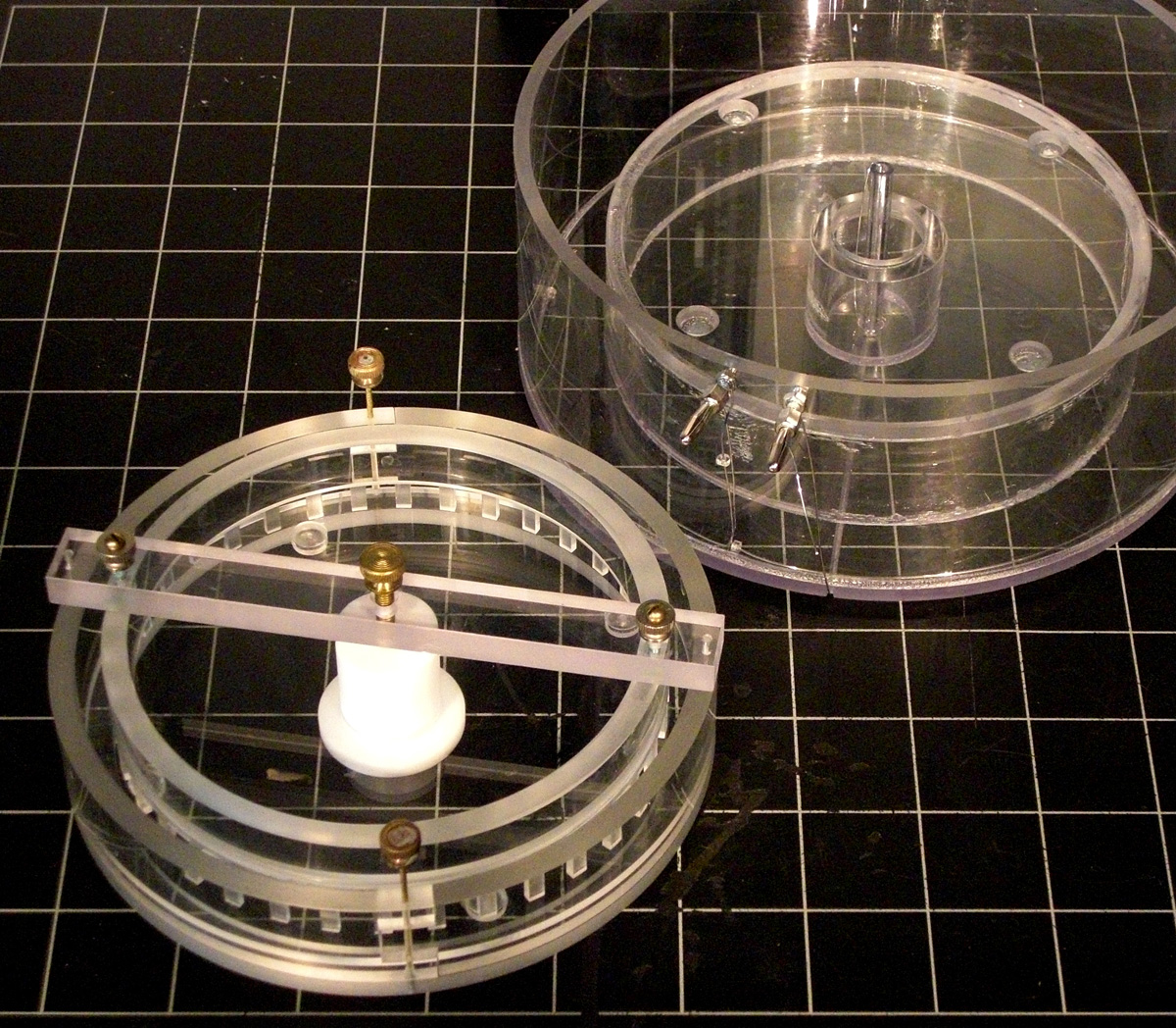
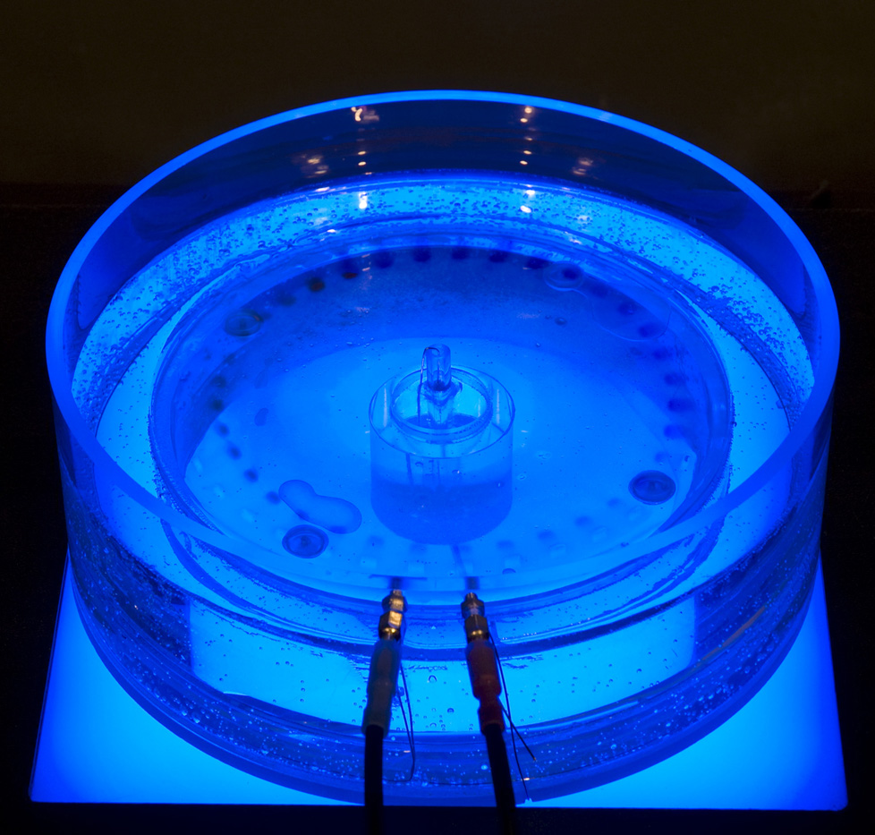
Ocular Revision, custom, circular electrophoresis rig shown.
Ocular Revision is an artwork incorporating an alternate mechanism for the analysis and display of the DNA image.  Typically, DNA is visualized in a rectangular chamber containing a porous gelatin that has an electrical field across it.  When DNA is inserted into one side of this gelatin the electrical current pulls it across the gel at a rate corresponding to its molecular mass and thus differentiates DNA of different sizes.

Ocular Revision uses a custom, experimental, circular gel electrophoresis rig to visualize DNA bands.  I designed this circular rig to be polarized from inside to outside of the circle.  The radial design and inside-out polarization allows the apparatus to create DNA images reminiscent not of a “barchart” or a “progress bar” on a computer screen, but rather a slow emergence; a signal; a flowering; an attraction or repulsion.  
Ocular Revision attempts to nudge DNA imaging back toward the realm of biology.  The goal (at least at present) is to force DNA to be read as substance rather than mere code and thus hopefully break a certain deadlock in Genetics caused by its overly simplistic operationalization.

The first images that I am creating with the 
circular DNA electrophoresis rigs are based upon hemispherical maps of the world.
These “Genetic Maps” could be interpreted as simplistic form-based puns in which the circle is a visual metaphor for a heavenly body like the earth.  But at a deeper level they call attention to ingrained metaphors such as “genetic mapping”, which are problematic because “mapping” implies (distanced) simplification, abstraction, and exploitation (i.e. political, and economic maps of the world).

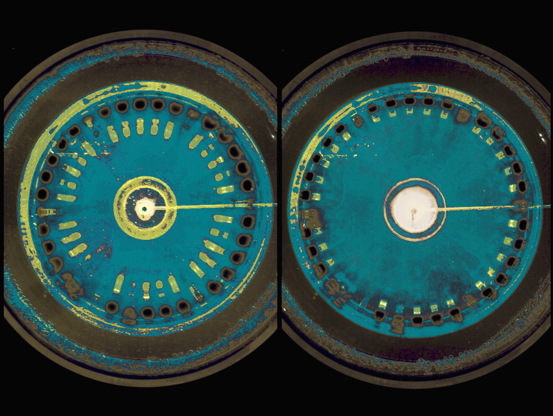
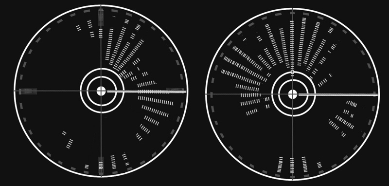
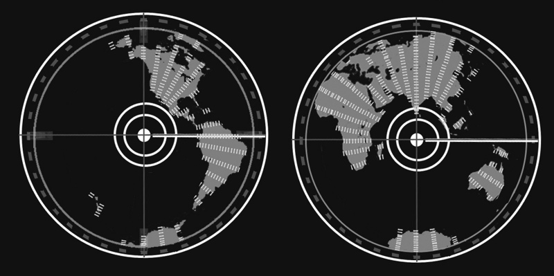
The three adjacent image pairs show:
(top) Actual human DNA imaged on the Ocular Revision circular electrophoresis aparatus.  Image formed is just a test to see if the device would work, not meant to form a specific image.
(middle and bottom) Sketches, hypothetically showing how the hemisheres of the earth might be produced from apropriately sized DNA fragments visualized in the Ocular Revision aparatus.

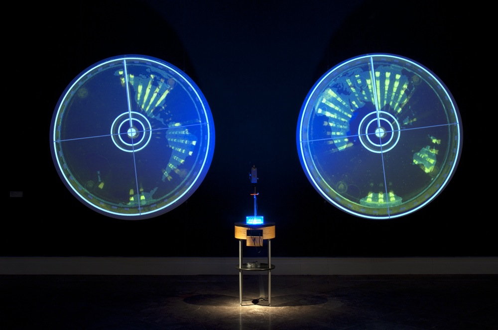
The final exhibition form of Ocular Revision is variable.  It will probably entail large images of the circular maps, generally these images will be time-based, perhaps they will be produced live, perhaps they will be accompanied by the circular electrophoresis apparatus itself.  See some of the first iterations below.


Ocular Revision at Surveyor, Albright-Knox Art Gallery, Buffalo:  the cicular cabinet contains the electrophoresis rig, camera, power supply and blue-light transilluminator.  Images of evolving map image are shown on suspended circular projection screens.  (photo by Tom Loonan.)


Ocular Revision at Biotopia: Art in the Wetzone, Aalborg, Denmark.  Close-up.
 

SUPPORTED BY:
Renew Media (Rockefeller Foundation), Media Arts Fellowship, 2008.
Prix ARS Electronica, Award of Distinction, Hybrid Arts, 2010.
VIDA 13.2, International Competition on Art and Artificial Life, Spain, Second Prize, 2011.


SPECIAL THANKS:
Sullivan Kerry Sheehan
Dr. Gerald Koudelka, University at Buffalo.


ADDITIONAL THANKS:
Dr. Helena Storchova, Institute of Experimental Botany, Prague.
Kevin Cullinan, University at Buffalo
Nathaniel Hall


VIDEO DOCUMENATION


Back to Vanouse projects